Left Shoulder Anatomy Diagram : MRI shoulder anatomy | shoulder coronal anatomy | free ... : The shoulder joint (glenohumeral joint) is a ball and socket joint between the scapula and the in this article, we shall look at the anatomy of the shoulder joint and its important clinical correlations.
byAdmin•
0
Left Shoulder Anatomy Diagram : MRI shoulder anatomy | shoulder coronal anatomy | free ... : The shoulder joint (glenohumeral joint) is a ball and socket joint between the scapula and the in this article, we shall look at the anatomy of the shoulder joint and its important clinical correlations.. Bone, then ligaments of the joint capsule, with tendons and muscles on top. Required fields are marked *. Last update february 25, 2021. Radiologists primarily perform shoulder imaging to assess injuries within the shoulder joint. Did not undergo a x ray or.
Posted on december 13, 2018december 12, 2018. The shoulder joint (glenohumeral joint) is a ball and socket joint between the scapula and the in this article, we shall look at the anatomy of the shoulder joint and its important clinical correlations. Radiologists primarily perform shoulder imaging to assess injuries within the shoulder joint. Related posts of diagram of shoulder muscles and tendons. Bone, then ligaments of the joint capsule, with tendons and muscles on top.
Shoulder Joint Diagram — UNTPIKAPPS from www.untpikapps.com This mri shoulder axial cross sectional anatomy tool is absolutely free to use. Starting with what is deepest, it goes: Remote distance is left up to 500m. Editor · aug 6, 2017 ·. 7 draw labelled diagram showing the relations of shoulder joint. Human anatomy for muscle, reproductive, and skeleton. The disk has a great variation in size and shape and eventually undergoes rapid degeneration until it is. Start studying shoulder anatomy diagram.
Did not undergo a x ray or.
We added an horizontal menu at. Movements of the human shoulder represent the result of a complex dynamic interplay of structural bony anatomy and biomechanics, static a thorough understanding of the functional anatomy of the shoulder provides the clinician with a foundation for caring for athletes with shoulder injuries. Leave a reply cancel reply. Radiologists primarily perform shoulder imaging to assess injuries within the shoulder joint. Webmd's shoulder anatomy page provides an image of the parts of the shoulder and describes its function, shoulder problems, and more. Normal anatomy, variants and checklist. The transverse humeral ligament is not shown on this diagram. 7 draw labelled diagram showing the relations of shoulder joint. Editor · aug 6, 2017 ·. .anatomy of shoulder high resolution wallpaper photos anatomy of shoulder bones shoulder joint ligaments pain on top of shoulder. Use the mouse scroll wheel to move the images up and down alternatively use the tiny arrows (>>) on both side of the image to move the images. Last update february 25, 2021. Shoulder anatomy diagram / normal shoulder anatomy.
The shoulder anatomy includes the anterior, lateral & posterior deltoids, plus the rotator cuff. The shoulder joint is encapsulated by a group of muscles and ligaments called the rotator cuff. We added an horizontal menu at. 8 name the arteries and the nerves that supply shoulder leave a reply cancel reply. Shoulder radiology & anatomy at usuhs.mil.
Bones of the shoulder region 3 - Wikiversity from upload.wikimedia.org The sagittal suture is the line where the right and left parietal bone are in contact. Posted on december 13, 2018december 12, 2018. Bone, then ligaments of the joint capsule, with tendons and muscles on top. Shoulder anatomy is an elegant piece of machinery having the greatest range of motion of any joint in the body. The shoulder is one of the largest and most complex joints in the body. 7 draw labelled diagram showing the relations of shoulder joint. .anatomy of shoulder high resolution wallpaper photos anatomy of shoulder bones shoulder joint ligaments pain on top of shoulder. 7 draw labelled diagram showing the relations of shoulder joint.
Use the mouse scroll wheel to move the images up and down alternatively use the tiny arrows (>>) on both side of the image to move the images.
As the disease progresses, night pain becomes more common. This is because the deltoids are what you would consider the major muscles of the shoulder anatomy; Remote distance is left up to 500m. To keep things simple, we can divide the shoulder into layers. This webpage presents the anatomical structures found on shoulder mri. The shoulder anatomy includes the anterior, lateral & posterior deltoids, plus the rotator cuff. The sagittal suture is the line where the right and left parietal bone are in contact. This acts as the bony framework by which the muscles of the chest, upper back and shoulder connect the upper limb to the trunk of the body and control it's movements.the clavicle connects to the sternum via the. The shoulder joint is the connection between the chest and the upper extremity. In this episode of eorthopodtv, orthopaedic surgeon randale c. The shoulder is one of the largest and most complex joints in the body. Human anatomical atlas of the shoulder : Related posts of diagram of shoulder muscles and tendons.
The sagittal suture is the line where the right and left parietal bone are in contact. Assessment | biopsychology | comparative | cognitive | developmental | language | individual differences | personality | philosophy | social | methods | statistics | clinical | educational | industrial | professional items | world psychology |. This acts as the bony framework by which the muscles of the chest, upper back and shoulder connect the upper limb to the trunk of the body and control it's movements.the clavicle connects to the sternum via the. Leave a reply cancel reply. The shoulder joint is formed where the humerus (upper arm bone) fits into the scapula.
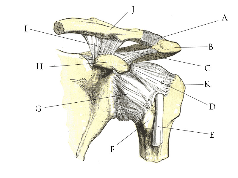
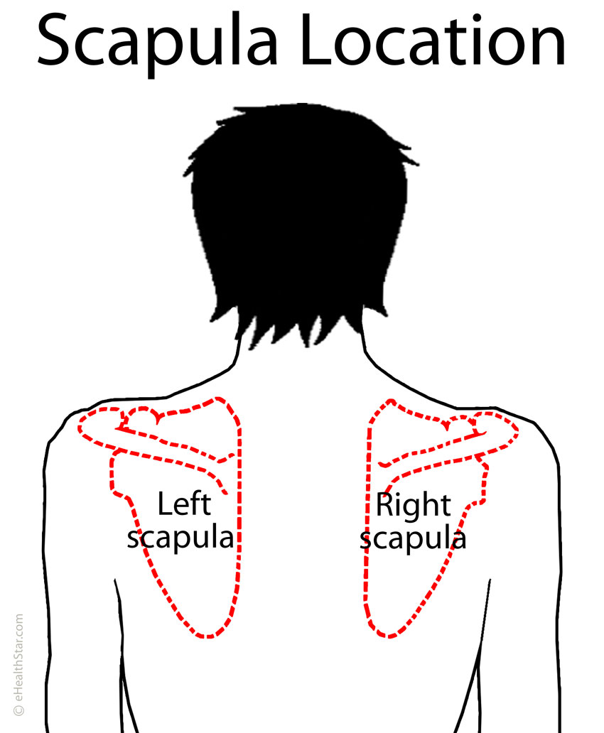
Scapula (Shoulder Blade) Anatomy, Muscles, Location ... from www.ehealthstar.com The clavicle (collarbone), the scapula (shoulder blade), and the humerus (upper arm bone) as well as associated muscles, ligaments and tendons. Bone, then ligaments of the joint capsule, with tendons and muscles on top. Learn vocabulary, terms and more with flashcards, games and other study tools. This acts as the bony framework by which the muscles of the chest, upper back and shoulder connect the upper limb to the trunk of the body and control it's movements.the clavicle connects to the sternum via the. The transverse humeral ligament is not shown on this diagram. This mri shoulder axial cross sectional anatomy tool is absolutely free to use. Movements of the human shoulder represent the result of a complex dynamic interplay of structural bony anatomy and biomechanics, static a thorough understanding of the functional anatomy of the shoulder provides the clinician with a foundation for caring for athletes with shoulder injuries. Instant anatomy is a specialised web site for you to learn all about human anatomy of the body with diagrams, podcasts and revision questions.
The human shoulder is made up of three bones:
Bone, then ligaments of the joint capsule, with tendons and muscles on top. Humbert's joint implant of my left hip enabled me to continue my love of hiking my favorite place. The shoulder joint is encapsulated by a group of muscles and ligaments called the rotator cuff. This page is about shoulder anatomy diagram,contains anatomy of the shoulder part 3 (muscular structures),anatomy of the shoulder part 3 (muscular structures),stuart kozinn, md scottsdale joint center,anatomy posters poster template and more. Leave a reply cancel reply. Click on a view to review its anatomy. Radiologists primarily perform shoulder imaging to assess injuries within the shoulder joint. Posted on december 13, 2018december 12, 2018. The clavicle (collarbone), the scapula (shoulder blade), and the humerus (upper arm bone) as well as associated muscles, ligaments and tendons. The human shoulder is made up of three bones: Understanding how the different layers of the shoulder are built and connected can help you understand how the shoulder works, how it can be injured, and how challenging recovery can be. The shoulder joint is formed where the humerus (upper arm bone) fits into the scapula. Use the mouse scroll wheel to move the images up and down alternatively use the tiny arrows (>>) on both side of the image to move the images.